
ENGINEERING · IMAGING · ARTIFICIAL INTELLIGENCE

Build technology around the gaps in care.

From automated microscopy to retinal imaging, microscopIA brings engineering and clinical expertise together to study tools that could expand access to specialist capacity.

Two programs. A shared purpose.

From a clinical need to evidence

  1. 01Define the unmet need
  2. 02Engineer and test
  3. 03Validate independently
  4. 04Assess readiness for use

Technology evidence · human-led by design

Devices and AI under development.

This evidence family documents concepts, physical prototypes, task-specific development benchmarks, validation gaps and human decision boundaries. microscopIA develops technology for settings where specialist capacity is limited without presenting investigational systems as clinical products.

CAD concept walkthroughSilent 45-second rotation of an early investigational CAD enclosure and base. It does not demonstrate clinical performance or an approved medical device.
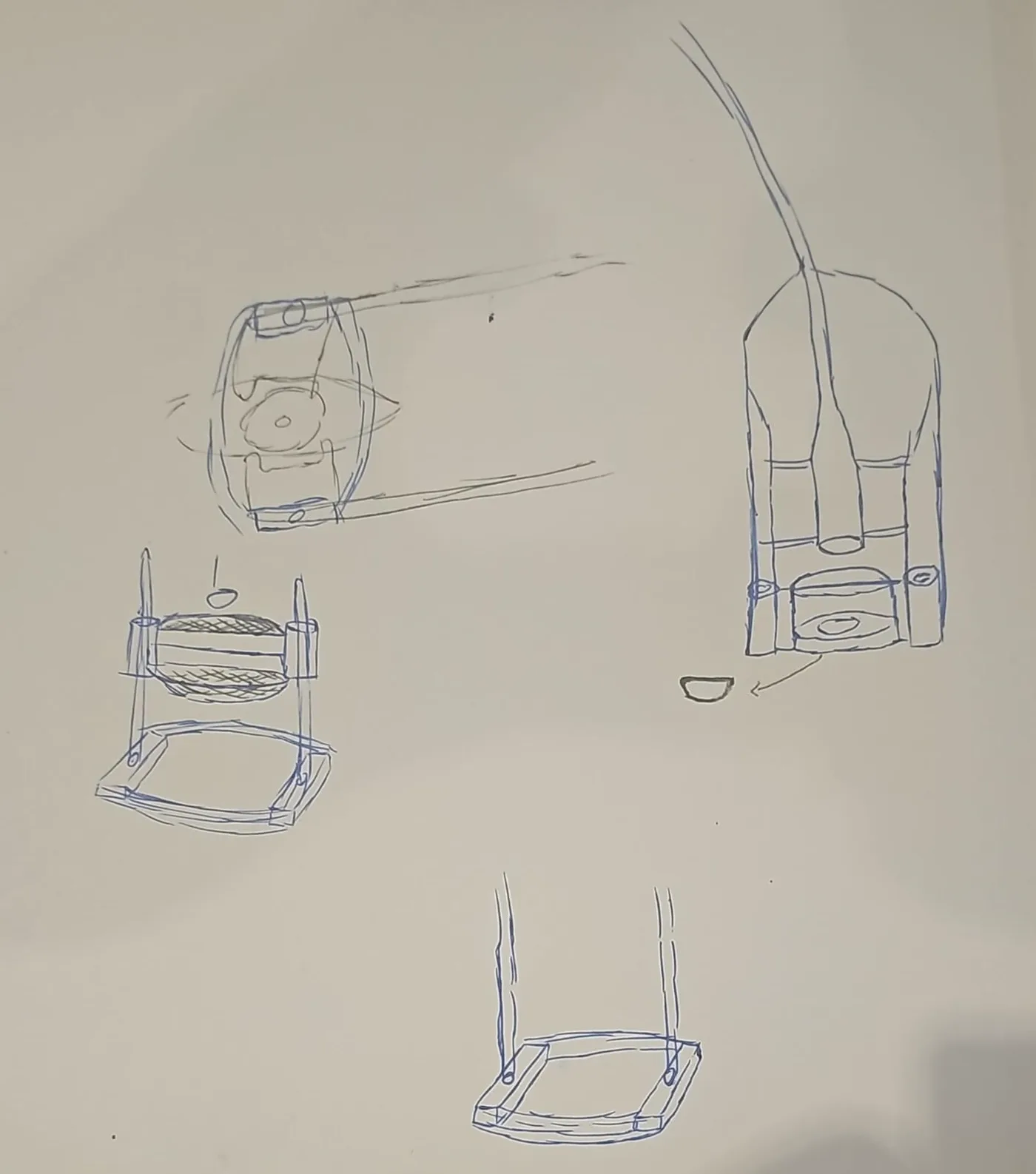
Hand-drawn early concept of a camera-support frame with adjustable supports and an examination base, shown from several angles.
Early concept sketchHand-drawn early concept of a camera-support frame with adjustable supports and an examination base, shown from several angles.

microscopIA-led device–AI program · research stage

Retinal-imaging and referral-support research for preterm infants

microscopIA is developing an integrated research system for retinal imaging in preterm infants. The device is intended to support a high-resolution camera, primarily a compatible smartphone, to improve inspection and capture of infant retinal images.

Research models analyze de-identified retinal images to study image quality and features of retinopathy of prematurity (ROP). The intended goal is to help identify infants with concerning findings or higher risk for referral to a ROP-trained ophthalmologist, who retains diagnosis and treatment decisions. Future use is being evaluated for appropriately trained neonatologists, neonatal nurses and physicians, including general practitioners where permitted, working under approved institutional protocols and local scope-of-practice rules.

Human-led research pathway

  1. Existing eligibility and follow-up protocol
  2. Image acquisition by a trained professional
  3. Image-quality check and research AI analysis
  4. Qualified clinical review
  5. Established specialist referral and follow-up

Ungradable image or failed acquisition → repeat or escalate under the approved protocol. There is no AI-created ‘no referral’ pathway.

Latest supplied AI development record

Task-specific development benchmarks—not clinical performance.

Source last modified 19 November 2025. Values are shown by model task because the supplied record does not establish performance of an integrated device–AI system.

Stage-related image classification

92.03Accuracy · reported scoreGCViT · 1,099 public-dataset images

Plus-disease classification

93.10Accuracy · reported scoreResNet50 · public development dataset

Vessel segmentation

76.81%Intersection over UnionU-Net · HVDROPDB
Methods, additional scores and limitations
Stage-related classification
Precision 93.39; recall/sensitivity 92.03; F1 92.32. Classes were Normal, Stages 1–3 and laser scars. Stages 4 and 5 were absent, so these results cannot support safe referral prioritization or deprioritisation.
Plus-disease classification
Precision 88.9; recall/sensitivity 89.8; F1 89.4. Dataset size, patient-level split and confidence intervals were not supplied.
Segmentation
IoU 76.81% using HVDROPDB and U-Net. The supplied record does not document external validation.
Not yet evidenced
The proposed binary ROP route has no supplied benchmark. Zone classification was not trained because the record reports that no suitable public dataset was identified.

The stage and plus values are reproduced as reported; the source does not print a percent sign for them. These are separate public-dataset development tasks with incomplete evaluation details. They are not directly comparable, externally or clinically validated, or evidence of device-level performance. No specificity, AUROC or confidence intervals are claimed.

Public video of the physical automated-microscopy research prototype.

microscopIA-led engineering · benchtop research prototype

Automated microscopy research for malaria

The public video documents a physical microscopy prototype. The program investigates automated image acquisition and software-assisted flagging of candidate fields, quality issues or samples for review by trained laboratory professionals. It does not confirm or exclude malaria, establish species or parasite density, choose treatment, or replace laboratory quality assurance and clinical judgement.

Any future application to another microscopy-based tropical disease requires its own intended use, dataset, validation plan and regulatory assessment.

Access by design

Research priorities start with settings where specialist people and infrastructure are scarce.

Human authority

Technology may support acquisition and prioritization; qualified professionals remain responsible for decisions.

Evidence before deployment

Safety engineering, usability, governance, training and independent validation precede clinical use.

Collaboration areas: ROP and pediatric ophthalmology, neonatology, optics and imaging, human factors, device engineering, clinical-AI validation, laboratory microscopy, quality systems and regulation.

Explore responsible funding

Development is not clinical authorization.

The work shown here is investigational. Qualified professionals retain clinical decisions. Each system needs evidence appropriate to its intended use, safety assessment and applicable regulatory review before clinical deployment.

Scientific and technology governance →